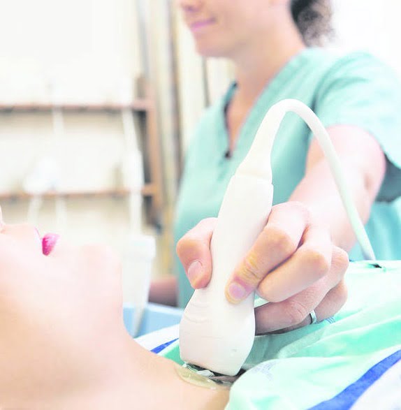
Knee, foot and ankle scan in London.
Knee problems and knee pain are common as the knee is a common point of contact in traumatic accidents and is prone to wear due to its stressful nature. It is also a common site for arthritic pain. The knee joint consists of four main structures: bone, cartilage, ligaments and tendons. The three bones come together to form the knee joint: the femur (femur), the tibia (tibia), and the patella (patella). The knee joint is made up of bones, cartilage, ligaments and tendons.
Ultrasound provides useful information on a wide range of conditions affecting parts of the knee, such as tendons, ligaments, muscles, synovial space, articular cartilage, and surrounding soft tissues. It may also rule out / confirm a Baker’s cyst on the back of the knee. The ankle and foot have many tendons, ligaments, and bones, pain or trauma are common causes to investigate the causes of tendinitis, bursitis, and degenerative changes such as arthritis, and joint effusion. Nodules such as ganglia, neuromas, and fibromas can be examined. Painful heels can be scanned to rule out / confirm plantar fasciitis.
What is the purpose of the knee examination?
The purpose of this ultrasound of the knee joint is to assess the main musculoskeletal structures of the knee joint. They include:
- Quadriceps tendon
- Patellar tendons
- Bursa
- Biceps femoris
- Anterior cruciate ligament
- Medial collateral ligament
- Lateral ligament
- Posterior cruciate ligament
Why do knee ultrasound?
- Pain
- Reduced motion
- Cancer and neoplasms
- Discomfort
- Inflammation
- Patellar tendon
- Patellar tendon rupture
- Quadriceps tendon rupture
- Prepatellar bursitis
- Subpatellar bursitis
- Popliteal cyst (Baker’s cyst)
- Broken kneecap
- A torn meniscus
- Broken ligament
- Torn hamstring muscle
- Gout (a form of arthritis)
Sports and overload injuries of the ankle and foot are common, and ultrasound has been recognized as an excellent diagnosis of foot and ankle pathology, providing a quick non-invasive research tool that is well tolerated by a patient with acute or chronic pain. The possibility of dynamic examination is another advantage of ultrasound in the assessment of pathology of the ankle and foot, where maneuvers such as muscle spasm and joint load can be particularly helpful.
What is the purpose of the ankle and ankle ultrasound?
The purpose of ankle ultrasound is to evaluate the ankle joint:
- Tendons
- Tendon sheaths
- Anterior articular space
- Calcaneus of the calcaneus
- Ligaments
Reasons for taking an ultrasound scan of the ankle and foot include:
- Pain
- Intra-articular bodies
- Rupture
- Tendon sheath inflammation
- Tendinitis
- Soft tissue masses
- Inflammation of the bursa or joint capsule
- Ligament injuries
- Effusion
- Ganglion cysts
- Plantar fasciitis
- Plantar fibroma
- Morton’s neuroma
- Cancer
- Foreign body
- Soft tissue masses such as ganglia, lipomas
- Tendinitis or tendon rupture
- Heel spurs
- Tarsal syndrome
- Muscle damage (chronic and acute)
- Joint exudates
- Vascular pathology
- Hematomas
- Mass classification, e.g. solid, cystic, mixed
- Postoperative complications, e.g. abscess, swelling

