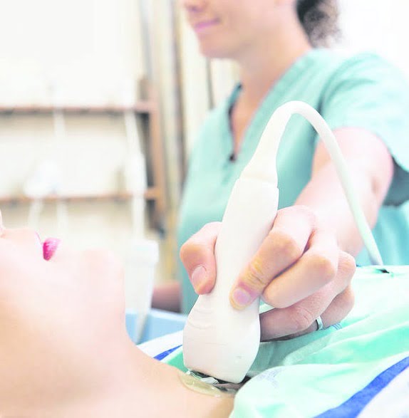
The ultrasound of the testicles in London is performed by a radiologist.
The study assesses, inter alia, testicles, epididymis, seminal cord, skin and subcutaneous tissue of the scrotum. Such examination should be made in the case of a palpable, painless thickening within the testicle, which will enable early detection of changes and assess their nature – benign or malignant neoplastic changes.
A testicular ultrasound is a diagnostic test that obtains images of the testicles and surrounding tissue in the scrotum. The two testicles are the main male reproductive organs. They produce sperm and the male sex hormone testosterone. Your testicles are located in the scrotum, which is the fleshy bag of tissue that hangs under your penis. Ultrasound is a safe, painless and non-invasive procedure. The procedure uses high-frequency sound waves to create images of the organs inside the body.
Ultrasound uses a probe or transducer. This portable device transforms energy from one form to another. Pushes energy to the target part of the body with sweeping movements. The transducer emits sound waves as it moves across your body. The transducer then picks up the sound waves that bounce off the organs in a series of echoes. The computer converts echoes into images on a video monitor. Normal and abnormal tissue carry different types of echoes. The radiologist can interpret the echoes to distinguish between benign and malignant tumor types.
Call our reception today to book your consultation.

