Doppler ultrasound examination at Eskulap Clinic London.

Doppler – how to prepare for the procedure?

Doppler ultrasonography of the carotid arteries is a very safe and simple examination. You don’t need to prepare for a carotid ultrasound at all, but wearing loose fitting clothes to keep you comfortable can help. During the scan, you will need to lie still for about half an hour. You will also need to expose your neck so that the ultrasound can make contact with the skin above the carotid arteries.

Enter your phone number and our consultant will call you back …

    What does the Doppler procedure look like?

    At the start of the test, a small amount of gel will be spread over the skin on both sides of the neck. This helps the ultrasonic probe to get a clearer image. The transducer will then be passed over your neck to take pictures of your carotid arteries. There are two arteries, each on either side of the neck. The transducer is a handheld probe that sends ultrasonic waves. It records bouncing echoes and converts them into images. The scan can create pictures of your arteries that show any narrowing or blockage. The Doppler ultrasound technique can also assess blood flow through the arteries by observing how the blood cells move. It allows the doctor to assess blood flow in real time.

    You may feel a slight pressure as the ultrasound probe is passed over your neck. The gel can also feel a bit cold when it is spread over the skin. However, Doppler examination of the carotid arteries is completely safe and painless. This can be a very effective way to assess your risk of stroke.

    Advantages of the ultrasound Doppler of the carotid arteries.

    • Ultrasound to exclude pathologies of the carotid arteries
    • A referral from a GP is not required
    • Same-day digital results
    • Fully qualified, experienced radiologists
    • No preparation is required
    • Same-day appointments are usually available.

    Aesthetic Medicine Skin Clinic in London and Birmingham office

    Find a treatment suitable for you … Type in your concern…

    Generic selectors
    Exact matches only
    Search in title
    Search in content
    Post Type Selectors
    Search in posts
    Search in pages
    Filter by Categories
    Uncategorized

    Enter your phone number and our consultant will call you back …

      Doppler of arteries and veins

      This test uses sound waves to show an ultrasound image of the structure of an artery and the amount of blood flowing through it. A gel is applied to the skin and a probe is placed over the area. Duplex ultrasound helps you determine where the narrow or blockage is in your arteries and how much it is narrowed. Obtaining a picture can be difficult if you are seriously overweight or if you have severe atherosclerosis. This test can help you decide which treatment is best for you.

      Risk of developing diseases of the heart and veins.

      • After insertion of the catheter, a pacemaker through a vein in the groin
      • Prolonged bed rest
      • Smoking
      • Family history of blood clots, embolism, and bleeding disorders
      • Fractures of the pelvis or limbs
      • Childbirth in the last 6 months
      • Heart failure
      • Obesity
      • Recent surgery (especially hip, knee or pelvic surgery)
      • Too many blood cells produced by the bone marrow (polycythemia vera), which makes the blood thicker and slower than normal

      You’re also more likely to develop deep vein thrombosis if you have any of the following conditions:

      • Blood that is more likely to clot (hypercoagulability)
      • Cancer
      • Taking estrogens or birth control pills. The risk is even greater if you smoke.

      Deep vein thrombosis is most common in adults over 60 years of age, but can occur at any age. Sitting for long periods, such as when traveling, may also increase your risk of developing deep vein thrombosis. This is most likely when one or more of the risk factors listed above are also present.

      Indications for the Doppler procedure of arteries and veins.

      • Deep vein thrombosis (blood clot) in the arms or legs
      • Narrowing of the arteries that restrict the flow (atherosclerosis, usually the arteries of the legs)
      • Peripheral veins to assess varicose veins and venous reflux (insufficiency)
      • Upper thoracic outlet syndrome
      • Swollen legs or arms, sometimes caused by a blood clot
      • Pain in the legs after walking a certain distance is sometimes caused by a reduced arterial blood flow to the legs due to disease (intermittent claudication)
      • Varicose veins, phlebitis, pain in the limbs
      • Blood clots can form when something slows down or changes the blood flow in the veins

      Assessment of abdominal aortic aneurysms.

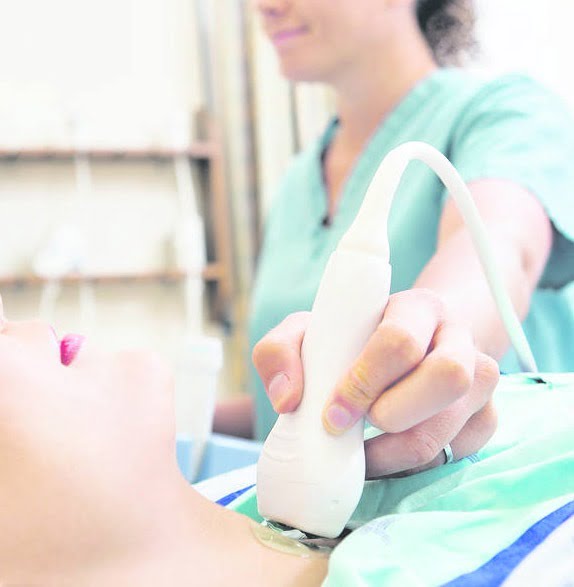
      Ultrasound is regularly used to assess abdominal aortic aneurysms. It is an enlargement of the aorta, the main blood vessel that supplies blood from the heart to the rest of the body. The aorta is about 2.5 cm around the abdomen, but it can reach a size of over 5 cm.

      Aneurysms are more common in men over the age of 65 than in women and younger men, and can be fatal as large aneurysms can rupture. According to NHS Choices, 8 out of 10 people with an aortic aneurysm die.

      Small aneurysms are very unlikely to cause any symptoms, but a large aneurysm can cause the following symptoms:

      • Intense pain in your back or stomach
      • Fast pulse
      • Nausea and vomiting
      • Excessive sweating
      • Although the causes are not fully understood, risk factors include obesity, high blood pressure, smoking, and high cholesterol

      Abdominal aorta scan includes the assessment of:

      • Abdominal aorta
      • Hip vessels

      Reasons for performing an ultrasound of the abdominal aorta:

      • Intense pain in your back or stomach
      • Pulsation, a ripple in the center of the abdomen
      • Fast pulse
      • Nausea and vomiting
      • Excessive sweating
      • Abdominal pain after a meal

      Vascular ultrasound

      Vascular ultrasound is performed to monitor blood flow to organs and tissues throughout the body, locating and identifying blockages and abnormalities such as blood clots or blockages.

      Vascular ultrasound can also help identify areas of abnormal vasodilation (aneurysm) that, if left untreated, could lead to serious consequences.

      A Doppler ultrasound scan can be part of an overall ultrasound scan. Doppler is a special ultrasound technique that assesses the speed and volume of blood flowing through a blood vessel, including the major arteries and veins in the abdomen, arms, legs, and neck. It can help diagnose blood flow blockages (such as clots, tumors, developmental anomalies), vasoconstriction (possibly caused by atherosclerosis), and tumors and birth defects.

      The images are captured in real time, so this is a dynamic exam and the exam recording can help your doctor monitor blood flow to organs and tissues throughout the body.

      How much does private ultrasound cost?

      At the Eskulap Clinic in London, we believe that private healthcare should be available to all of us. That is why we always try to keep the costs of our ultrasound services as low as possible, without compromising the quality of care. All research prices are listed in the price list, with no hidden costs or unpleasant surprises.

      Contact data

      London:
      147 Ruislip Road,
      Greenford, London
      UB6 9QQ

      Birmingham:
      Shakespear Crescent
      Hockley, Birmingham
      B18 5BT

      phone 07747 474755

      Opening hours:

      Mon – Sat 11:00 – 18:00

      Generic selectors
      Exact matches only
      Search in title
      Search in content
      Post Type Selectors
      Search in posts
      Search in pages
      Filter by Categories
      Uncategorized

      Contact Form